Introduction
Toad is a common name for certain frogs, amphibians that are characterized by dry, leathery skin, short legs, and large bumps covering the parotid glands. Toads are especially of the family Bufonidae. The family Bufonidae contains about 500 species which includes the Stream Toads, Cape Toads, Tree Toads, Dwarf Toads, Common African Toads, etc. The Common African Toad is the case study of this practical.
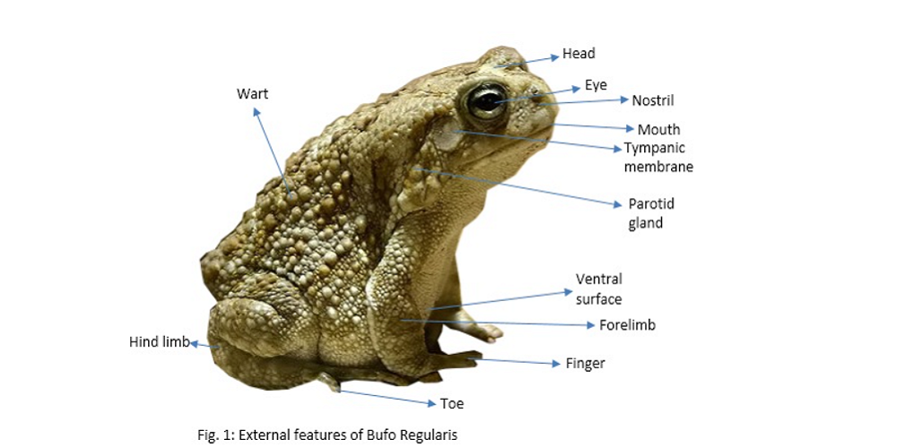
The Common African Toad is known as Bufo regularis. It is a specie toad in the family Bufonidae; and found widely in the Sub-Saharan Africa. The external features are as shown in figure 1. Note that the body is divided into head and a trunk which bears a pair of short fore-limbs and a pair of long powerful hind-limbs. The head bears a wide terminal mouth, a pair of external nostrils; a pair of comparatively large eyes; tympanic membranes or eardrums located behind the eyes; and poison glands which are large oval patches of raised skin behind the tympanic membranes.
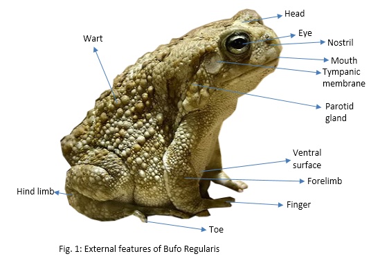
The trunk bears a cloacal opening at its posterior end, the paired forelimbs each terminating in four digits, and the hind-limbs in five, much longer webbed digits. All these digits possess tubercles or pads on their underside. To identify the sex of toads, it should be noted that the skin beneath the chin is dark in the male whereas it is creamy white in the female.
The toad as an amphibian is able to receive oxygen and give out carbon dioxide from three kinds of respiratory organs: the lungs, the skin, and the mucous membrane. For the pulmonary respiration, organs concerned include the internal and external nares, the glottis, the lungs, the bronchi, the laryngotracheal chamber, and the buccal cavity. The buccal cavity of the toad can be examined by opening its mouth. Notice that the tongue is attached to the floor anteriorly so that its free end is towards the throat. The floor’ of the mouth is supported by hyoid apparatus which is a cartilaginous skeleton. On this floor, there are two apertures behind the eyeball bulges and these are the Eustachian tubes which lead to the middle ear on each side in the male, beneath the tongue, there is another pair of slit like openings leading to the vocal sac. Right at the back, there is a slit like aperture, which is the glottis.
Dissection Process
1. Note that vertebrates are normally dissected from the ventral surface and invertebrates from the dorsal.
2. Pin the toad with its ventral side uppermost and limbs extend under water.
3. Lift the skin of the trunk with forceps and make a mid-ventral incision backward to the cloaca and forwards to the chin.
4. Make other incision from the mid-ventral line to the knees and the elbows on each side.
5. Pin out the flaps of the skin after separating it from the inner muscular wall. In the male, the vocal sac is seen as a dark membranous sac in front of the pectoral girdle and muscles which lie between the hind limbs. The viscera are enclosed within the muscular body wall between the pectoral and pelvic muscles.
6. Locate the anterior abdominal vein which shows through the ventral body wall towards the posterior end of the body.
7. On one side of the anterior abdominal vein, lift the body wall and make an incision. Continue this cut anteriorly through the pectoral girdle and posteriorly as far as possible. Make a similar cut on the other side of the anterior abdominal vein.
8. Very near the pelvic muscles, cut transversely through the muscular body wall carefully pull the strip towards freeing it from the underlying tissue. By so doing, the anterior abdominal vein is left intact. Cut the body transversely near the pectoral and pelvic girdles and pin back the two flaps of tissue so formed on each side of the body.
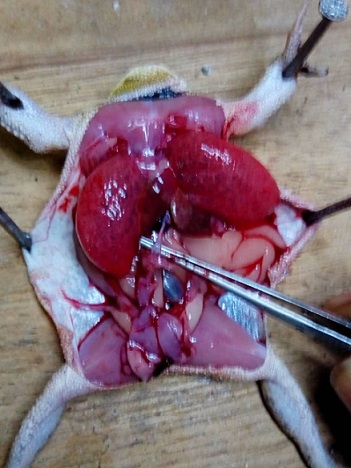
9. Identify the liver which is divided into right and left lobes, the gall, which is a green globular structure, the stomach, the duodenum, the small intestine, the pancreas lying in the loop made by the stomach and the duodenum, the rectum, the spleen which is spherical dark red structure and the fat bodies. The heart is covered by a thin membrane, the pericardium, and it consists of a ventricle, two auricles, and a sinus venosus. Dorsal to the liver are the lungs, a pair of thin-walled pink sacs.
10. Deflect the posterior region of the gut to the right and observe the kidneys and the cream-coloured testes which are dorsal to the small intestine and rectum.
11. In female, ovaries appear as large black masses on either side of the viscera and lying above kidneys. The urinary bladder is a whitish. thin-walled bilobed structure, and it lies at the posterior end eel the body.
12. Make a median incision with a scalpel through the pelvic girdle, leaving the anterior abdominal vein intact and part the hind limbs to expose the rectum.
13. Display the alimentary canal by pinning the stomach on the left side of the toad and the small intestine without tearing the mesenteries on the right.
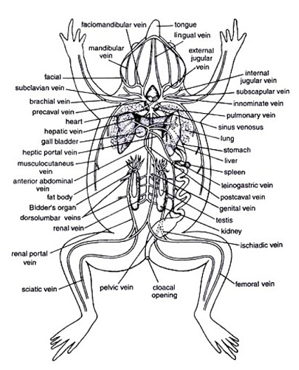
Result
A typical dissected view of a toad is as shown below:
References
1. ZOO 103: Experimental Zoology 1. ‘Class Amphibia’. Obafemi Awolowo University, Ile-Ife.
2. www.biologydiscussion.com. ‘Respiratory System in Toad (With Diagram) | Zoology’. Assessed on July 28, 2018.
3. en.wikipedia.org. ‘Toad’. Assessed on July 28, 2018.
4. en.wikipedia.org. ‘Amietophyrynus Regularis’ Assessed on July 28, 2018.


